Anatomy Of Upper Thigh And Hip : Hip Strains | Tight hips, Tight hip flexors, Hip anatomy / The following nerves serve the gluteal and thigh regions:. When walking on slick surfaces, pay attention to your steps. The ball is the rounded end of the femur (also called the. These are the piriformis, obturator internus, obturator externus, gemellus superior, gemellus inferior, and quadratus femoris. If the pain is located in the groin or in the thigh, this is known as psoas bursitis, which affects the muscle that connects the femur to the lumbar vertebrae. The pelvis and the femur (the thighbone).
Medial condyle of tibia nerve supply: Hip pain can also refer pain up to the lower back and down into the groin. Anatomy hip, thigh and leg muscles. They are also known as the inner hip muscles and deep external rotators. Differential diagnosis differential diagnosis of muscular upper leg and hip pain includes:
Keeping on Track with Knees | Expanding Light from www.expandinglight.org It is the largest bone in the body. Like the forearm, the upper leg, or thigh, has a dense arrangement of many muscles. The upper leg is often called the thigh. Differential diagnosis differential diagnosis of muscular upper leg and hip pain includes: The femur is the upper leg bone or thigh. Anatomy of the human body. B, muscles of the anterior thigh compartment. Iliac crest tilts forward in a sagittal plane;
Now that you watched the video, you shou.
The transverse axis permits flexion and extension movement. The abductor muscles perform the opposite function to the adductors, pulling your upper legs away from your midline. The location of the center of the entire axis is at the femoral head. The hip is formed where the thigh bone (femur) meets the three bones that make up the pelvis: Small and deep muscles which mainly externally rotate the thigh at the hip joint and stabilize the pelvis. The thigh is the region between the hip and knee joints. These are the piriformis, obturator internus, obturator externus, gemellus superior, gemellus inferior, and quadratus femoris. The following nerves serve the gluteal and thigh regions: Related online courses on physioplus. The femur, the hip bone (subdivided into ilium. The posterior muscle group is made up of the muscles that extend (straighten) the thigh at the hip. They are also known as the inner hip muscles and deep external rotators. The only bone in the thigh is the femur, which extends from the hip to the knee.
By adulthood, these three bones are completely fused and the pelvis is effectively a single bone. The rectus femoris is located in the center of the thigh, while the vastus medialis is in the middle of the said body part. Origins/insertions, actions and innervations learn with flashcards, games, and more — for free. When walking on slick surfaces, pay attention to your steps. The ilium, the pubis (pubic bone) and the ischium.
leg | Definition, Bones, Muscles, & Facts | Britannica from cdn.britannica.com Anatomy of the human body. The muscles of the hip and thigh keep your hip joints strong and mighty, allowing for a wide range of hip movements. Anatomy hip, thigh and leg muscles. Hip and leg pain can cause stress on joints and affect other areas of the body. The thigh is the region between the hip and knee joints. As group, these muscles act to extend at the hip, and flex at the knee. The hip is formed where the thigh bone (femur) meets the three bones that make up the pelvis: The thigh bone or femur and the pelvis join to form the hip joint.
The thigh muscles are divided into three compartments:
When walking on slick surfaces, pay attention to your steps. Like the forearm, the upper leg, or thigh, has a dense arrangement of many muscles. B, muscles of the anterior thigh compartment. Now that you watched the video, you shou. It's the area that runs from the hip to the knee in each leg. Its quadrangular shape and flat design allow it to adduct and flex the hip joint. This portion is referred to as the head of the femur, or femoral head. Muscles of the hip joint. Rectus femoris forms the middle portion of the quadriceps. The joints and muscles of the hips and thighs need nervous input so they can do what your brain wants them to do. These three bones converge to form the acetabulum, a deep socket on the outer edge of the pelvis. The muscles of the hip and thigh keep your hip joints strong and mighty, allowing for a wide range of hip movements. Small and deep muscles which mainly externally rotate the thigh at the hip joint and stabilize the pelvis.
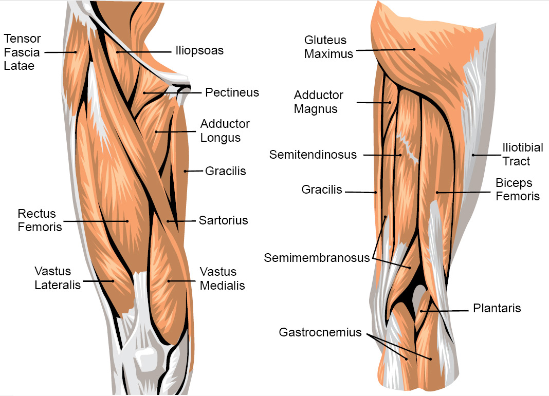
New headache, swollen hip, swollen hips, swelling of one hip, leg swelling, weakness of both legs, leg weakness On the anterior side, the most prominent of the muscles are the sartorius muscle and the four muscles that make up quadriceps muscle group (the quads.) The only bone in the thigh is the femur, which extends from the hip to the knee. Bony prominence on the proximal lateral side of the thigh, just below the hip joint. Meanwhile, the vastus lateralis is on the side of the thigh, while the vastus intermedius is hidden below the rectus femoris(5).
Hip Pain Associated Symptoms, Causes & Treatment from images.emedicinehealth.com People who play soccer have these specific muscles of the leg very well defined, so they're like a walking anatomy atlas for thigh muscles. The four muscles all extend the lower leg. B, muscles of the anterior thigh compartment. Muscles of the hip joint. The hip joint is made up of two bones: The joints and muscles of the hips and thighs need nervous input so they can do what your brain wants them to do. The femur is the upper leg bone or thigh. Related online courses on physioplus.
The pelvis and the femur (the thighbone).
Abductors are located on the upper portion of the outside of your thighs and hips, anchoring above on the pelvis, and below at various points on your outside thigh. Anatomy hip, thigh and leg muscles. The ilium, the pubis (pubic bone) and the ischium. Iliopsoas muscle, a hip flexor muscle that attaches to the upper thigh bone upper thigh anatomy. On the anterior side, the most prominent of the muscles are the sartorius muscle and the four muscles that make up quadriceps muscle group (the quads.) Next to the femoral neck, there are two protrusions known as greater and lesser trochanters which serve as sites of muscle attachment. Bony prominence on the proximal lateral side of the thigh, just below the hip joint. The location of the center of the entire axis is at the femoral head. The hip muscles are going to be slip into hip muscles and gluteal muscles. Ebraheim's educational animated video describes muscle anatomy of the thigh. Meanwhile, the vastus lateralis is on the side of the thigh, while the vastus intermedius is hidden below the rectus femoris(5). It is the largest bone in the body. The pelvis and the femur (the thighbone).
These are the piriformis, obturator internus, obturator externus, gemellus superior, gemellus inferior, and quadratus femoris upper thigh anatomy. At the top of the femur is a rounded protrusion which articulates with the pelvis.


0 Komentar