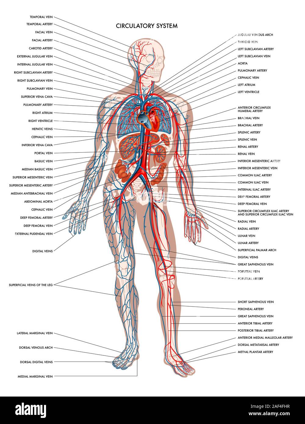
Abdominal Blood Vessels Labeled / Our purpose was to evaluate the location of the major blood vessels of the abdominal wall relative to landmarks apparent at laparoscopy.
Abdominal Blood Vessels Labeled / Our purpose was to evaluate the location of the major blood vessels of the abdominal wall relative to landmarks apparent at laparoscopy.. It then continues downward into the abdomen, where it branches into the iliac arteries just above the pelvis. This full color stock medical exhibit illustrates the normal anatomy of the abdominal blood vessels. Common incisions and closure techniques, and prevention and management of wound complications, are discussed elsewhere. As the abdomen and pelvis contain the majority of internal organs, these regions need to be supplied by an extensive network of arteries and veins. It begins at t12 and ends at l4 with its bifurcation into the common iliac arteries and usually has the following branches:
The aorta is the largest blood vessel in the body. Katy wallis at state college of florida In human anatomy, inferior epigastric artery refers to the artery that arises from the external iliac artery.it anastomoses with the superior epigastric artery.along its course, it is accompanied by a similarly named vein, the inferior epigastric vein.these epigastric vessels form the lateral border of hesselbach's triangle, which outlines the area through which direct inguinal hernias protrude. The abdominal aorta supplies blood to much of the abdominal cavity. The videos are done by dr.
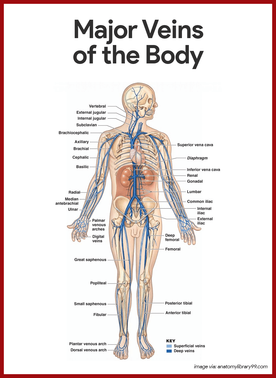
Blood Vessels Of Abdomen And Pelvis Anatomy Overview Kenhub from thumbor.kenhub.com Label the intestinal structures using the hints provided. The superior vena cava is the large vein that brings blood from the head and arms to the heart, and the inferior vena cava brings blood from the abdomen and legs into the heart. The aorta is the large artery leaving the heart. More posterior than the ivc until the umbilicus level where it lies more anterior than the ivc. 3 the superficial vessels include the superficial epigastric and the superficial circumflex iliac vessels. These vessels are branches of the femoral artery and vein. Abdominal wall anatomy that is clinically pertinent to the surgeon, focusing primarily on the structures of the anterior abdominal wall, will be reviewed. Understand the function of the thoracic and abdominal.
Label the abdominal contents using the hints provided.
Courses inferior through chest and enters abdomen through the diaphragm. Nodes drain to preaortic lymph nodes in root of primary arteries of gut (celiac nodes, superior and iferior mesenteric nodes) The abdominal aorta enters the abdomen through the diaphragm at the level of the twelfth thoracic vertebre and continues to just below the umbilical area, where it splits into the right and left common iliac arteries. Instant anatomy is a specialised web site for you to learn all about human anatomy of the body with diagrams, podcasts and revision questions Dissection of the blood vessels posterior to the diaphragm procedure: Katy wallis at state college of florida It has a number of important relationships and branches, which very commonly appear in exam questions and anatomy spotters. The abdominal aorta supplies blood to much of the abdominal cavity. Label the abdominal blood vessels using the hints provided. Note that the bifurcation (union) of the inferior vena cava is at l5 and therefore below that of the bifurcation of the aorta. Understand the function of the thoracic and abdominal. Common incisions and closure techniques, and prevention and management of wound complications, are discussed elsewhere. It begins at t12 and ends at l4 with its bifurcation into the common iliac arteries and usually has the following branches:
Common incisions and closure techniques, and prevention and management of wound complications, are discussed elsewhere. Courses inferior through chest and enters abdomen through the diaphragm. Katy wallis at state college of florida Instant anatomy is a specialised web site for you to learn all about human anatomy of the body with diagrams, podcasts and revision questions Nodes drain to preaortic lymph nodes in root of primary arteries of gut (celiac nodes, superior and iferior mesenteric nodes)
Circulatory System High Resolution Stock Photography And Images Alamy from c8.alamy.com 3 the superficial vessels include the superficial epigastric and the superficial circumflex iliac vessels. This full color stock medical exhibit illustrates the normal anatomy of the abdominal blood vessels. The abdominal aorta supplies blood to much of the abdominal cavity. These vessels are branches of the femoral artery and vein. Practice identifying the blood vessels on the photographs here and in your fetal pig photoalbum online. The videos are done by dr. It is an artery, meaning that it carries blood away from the heart. Advertising on our site helps support our mission.
Dissection of the blood vessels posterior to the diaphragm procedure:
Label the abdominal contents using the hints provided. The aorta begins at the left ventricle of the heart, extending upward into the chest to form an arch. This full color stock medical exhibit illustrates the normal anatomy of the abdominal blood vessels. Common incisions and closure techniques, and prevention and management of wound complications, are discussed elsewhere. 3 the superficial vessels include the superficial epigastric and the superficial circumflex iliac vessels. Nodes drain to preaortic lymph nodes in root of primary arteries of gut (celiac nodes, superior and iferior mesenteric nodes) More posterior than the ivc until the umbilicus level where it lies more anterior than the ivc. Advertising on our site helps support our mission. Aortas or aortae 4) is the main blood vessel in the abdominal cavity that transmits oxygenated blood from the thoracic cavity to the organs within the abdomen and to the lower limbs. Abdominal wall anatomy that is clinically pertinent to the surgeon, focusing primarily on the structures of the anterior abdominal wall, will be reviewed. Label the abdominal contents using the hints if provided. I hope this anatomy guide is helpful. It has a number of important relationships and branches, which very commonly appear in exam questions and anatomy spotters.
We will include an analysis of the normal doppler waveforms of the abdominal vessels. The common iliac arteries and veins. The identification of abdominal vessels using ultrasound is based on knowledge of their normal location, appearance and relationship to specific organs. Abdominal wall anatomy that is clinically pertinent to the surgeon, focusing primarily on the structures of the anterior abdominal wall, will be reviewed. This video series covers the blood vessels for anatomy and physiology ii students.
Cardiovascular System Anatomy And Physiology Study Guide For Nurses from nurseslabs.com Of course, recognition of the normal vascular anatomy is essential for the investigation of any abdominal vascular problem. Label the abdominal contents using the hints provided. This video series covers the blood vessels for anatomy and physiology ii students. Note that the bifurcation (union) of the inferior vena cava is at l5 and therefore below that of the bifurcation of the aorta. Label the abdominal contents using the hints if provided. Aorta (aorta) the aorta is the first segment of the systemic arterial circulation, originating directly from the left ventricle of the heart.it is the largest artery in the body consisting of three parts that each has its special characteristics, most notably in their direction and orientation. It then continues downward into the abdomen, where it branches into the iliac arteries just above the pelvis. The presence of vascular loops allows surgeons to ligate individual vessels with the expectation that blood will find its way to a particular region by alternate branches.
Advertising on our site helps support our mission.
Label the abdominal contents using the hints provided. Label the intestinal structures using the hints provided. Teachme anatomy part of the teachme series the medical information on this site is provided as an information resource only, and is not to be used or relied on for any diagnostic or treatment purposes. Doppler studies of the abdominal vessels demand an understanding of normal and abnormal blood flow patterns. The identification of abdominal vessels using ultrasound is based on knowledge of their normal location, appearance and relationship to specific organs. Courses inferior through chest and enters abdomen through the diaphragm. The aorta is the largest blood vessel in the body. Located anterior and to the left of the spine and to the left of the ivc. Common incisions and closure techniques, and prevention and management of wound complications, are discussed elsewhere. Lymph from abdominal viscera drains to lymphatic channesl and multiple named nodes alongisde arteries. The venous drainage of the abdomen is carried out by the portal venous system and the systemic venous system. Label the abdominal contents using the hints if provided. We will include an analysis of the normal doppler waveforms of the abdominal vessels.
Anatomy of blood vessels of abdomen pelvic cavities blood vessels labeled. This artery is responsible for transporting oxygen rich blood from your heart to the rest of your body.


0 Komentar